41 cell membrane diagram with labels
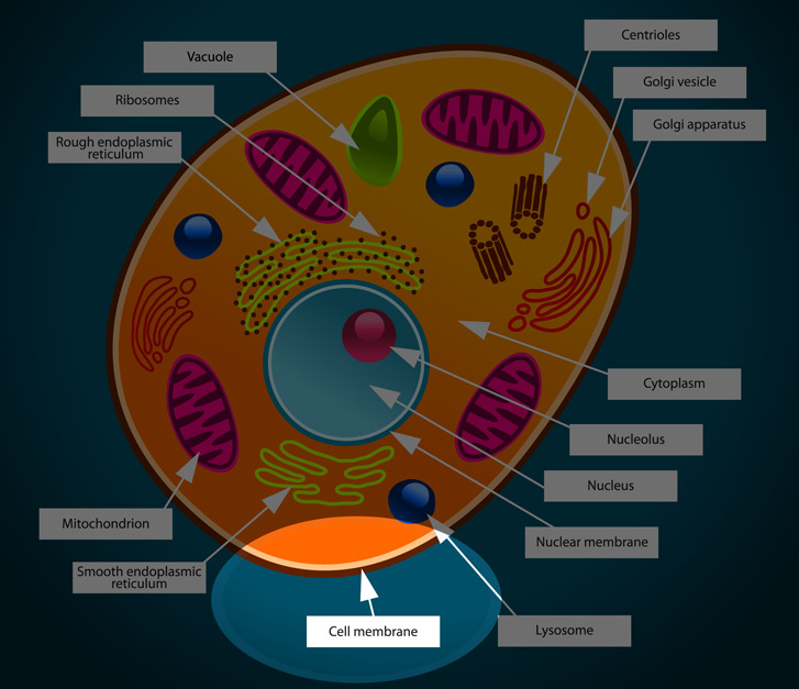
A Labeled Diagram of the Animal Cell and its Organelles A Labeled Diagram of the Animal Cell and its Organelles There are two types of cells - Prokaryotic and Eucaryotic. Eukaryotic cells are larger, more complex, and have evolved more recently than prokaryotes. Where, prokaryotes are just bacteria and archaea, eukaryotes are literally everything else. PDF Membrane Structure and Function - Phoenix College the cell membrane. Major Components of the Cell Membrane: Lipids • Phospholipids are amphipathic molecules (with hydrophobic tails and a hydrophilic head) • One of the phospholipid tails exist mostly in a trans configuration, providing more fluidity to the membrane • Cholesterol is a rigid
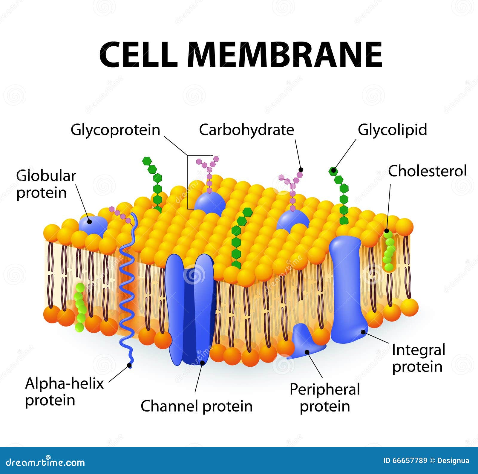
File:Cell membrane detailed diagram 4.svg - Wikipedia Cell membrane detailed diagram 4.svg. English: The cell membrane, also called the plasma membrane or plasmalemma, is a semipermeable lipid bilayer common to all living cells. It contains a variety of biological molecules, primarily proteins and lipids, which are involved in a vast array of cellular processes.
Cell membrane diagram with labels
Diagram of a cell membrane with labels - nist.gov Essential Biological FunctionsImmune response, Cell metabolism, Neurotransmission, Photosynthesis, Cell adherence, Cell growth and differentiationPotential Commercial ApplicationsDrug response monitoring, Chemical manufacturing, Biosensing, Energy conversion, Tissue engineering ... Diagram of a cell membrane with labels. Appears In. Biology in ... Cell Membrane - The Definitive Guide | Biology Dictionary The cell membrane, also known as the plasma membrane, is a double layer of lipids and proteins that surrounds a cell. It separates the cytoplasm (the contents of the cell) from the external environment. It is a feature of all cells, both prokaryotic and eukaryotic. a 3D diagram of the cell membrane Function of the Cell Membrane Cell: Structure and Functions (With Diagram) - Biology Discussion Eukaryotic Cells: 1. Eukaryotes are sophisticated cells with a well defined nucleus and cell organelles. 2. The cells are comparatively larger in size (10-100 μm). 3. Unicellular to multicellular in nature and evolved ~1 billion years ago. 4. The cell membrane is semipermeable and flexible. 5. These cells reproduce both asexually and sexually.
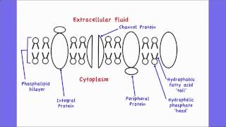
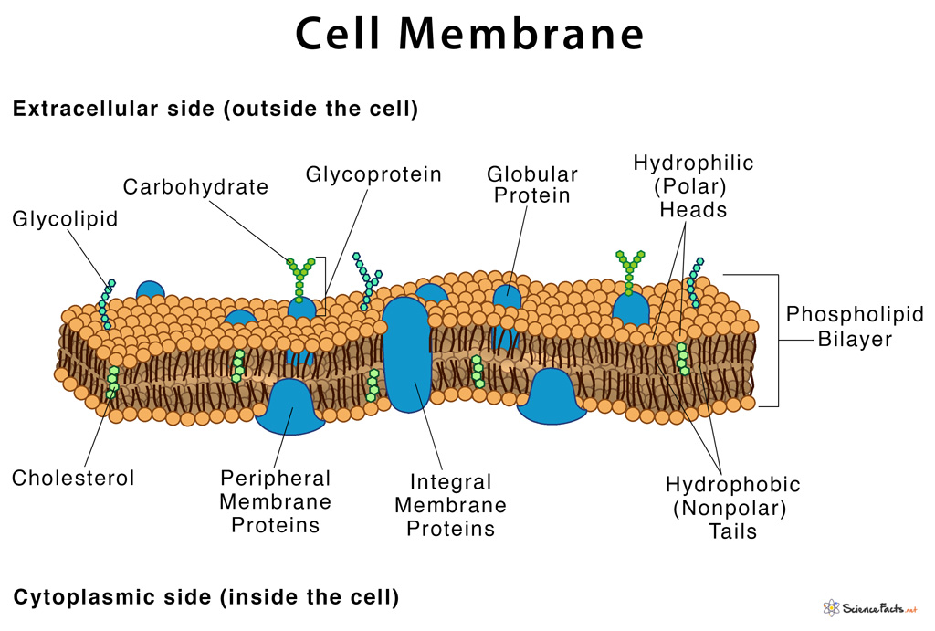
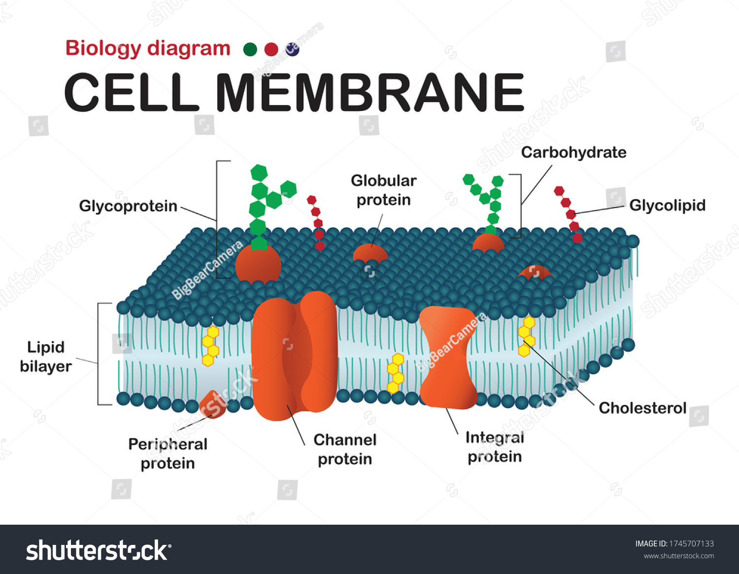
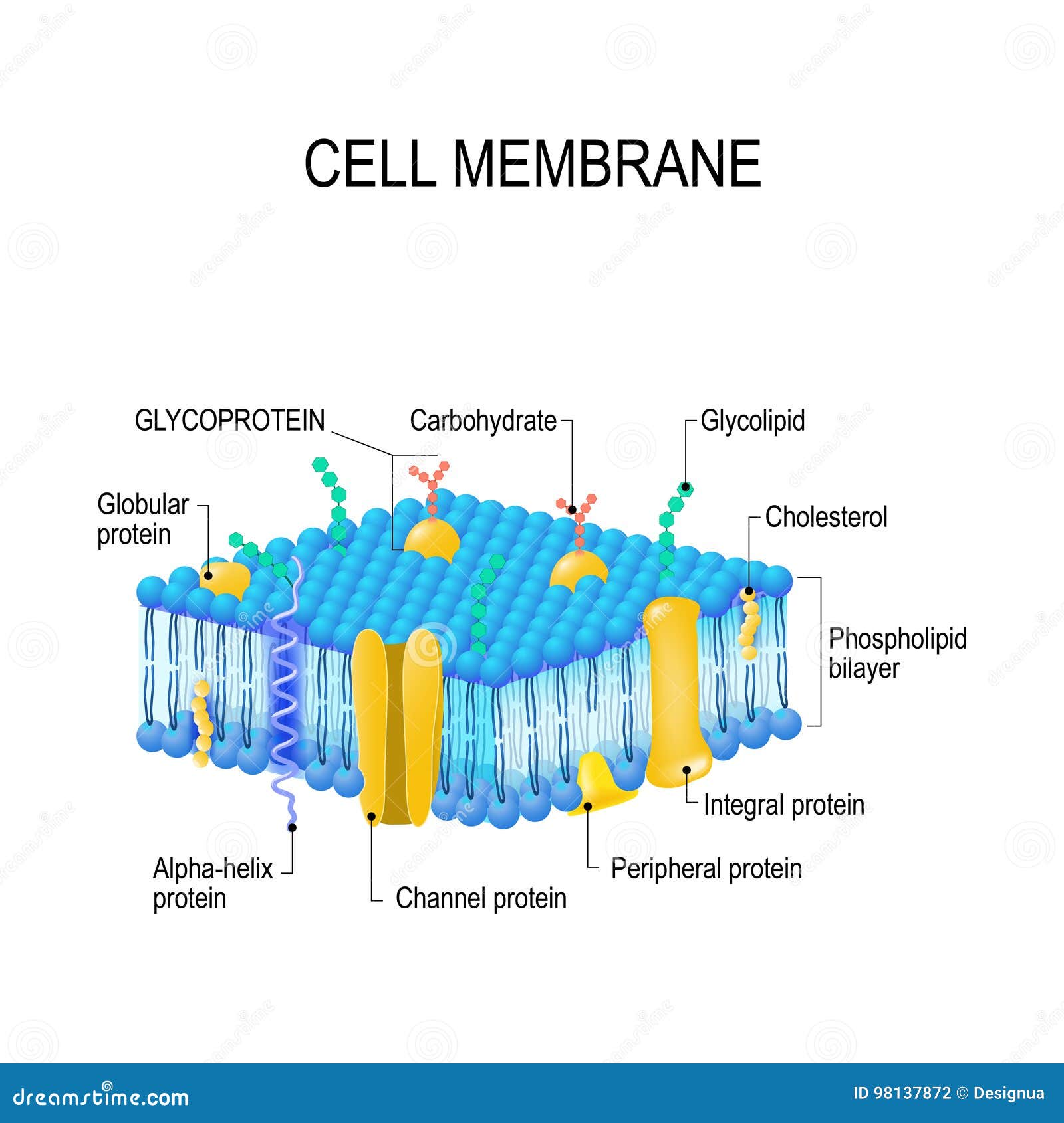
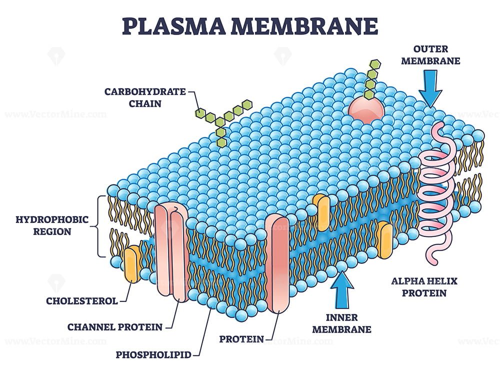
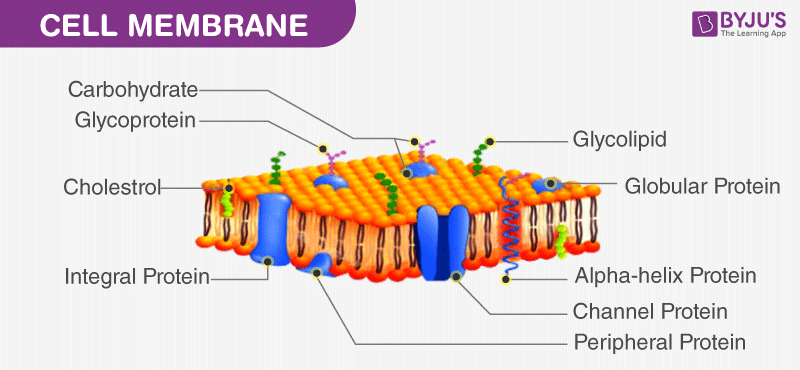
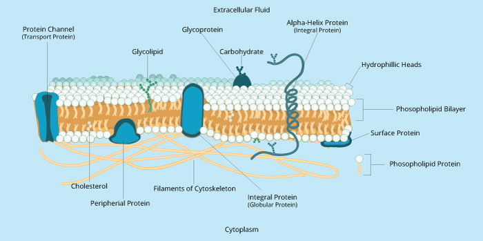
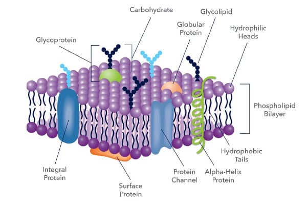
Cell membrane diagram with labels. Cell Membrane Labeling | Cell Structure Quiz - Quizizz Question 9. 30 seconds. Q. The function of carbohydrates in the cell membrane is to. answer choices. stick to other cells and sense stuff outside the cell. create energy for the cell. form the phospholipid bilayer. allow molecules to pass through it. Cell Membrane Labeled | EdrawMax Template Mar 2, 2022 ... In the cell membrane labeled diagram, we see Glycolipid, Glycoprotein, Globular Protein, Carbohydrate, Cholesterol, Peripheral Protein, ... Label the Cell Membrane - Labelled diagram - Wordwall Label the Cell Membrane - Labelled diagram. Home. Features. Contact. Price Plans. Log In. Sign Up. Language. channel protein, cholesterol, external cell environment, hydrophilic (water loving) part of phospholipid bilayer, peripheral protein, internal environment of the cell, hydrophobic (water fearing) part of phospholipid bilayer, glycolipid. THE PLASMA MEMBRANE. - Pinterest Feb 15, 2021 - THE QUESTION IS: Correctly label the following anatomical features of a plasma membrane.
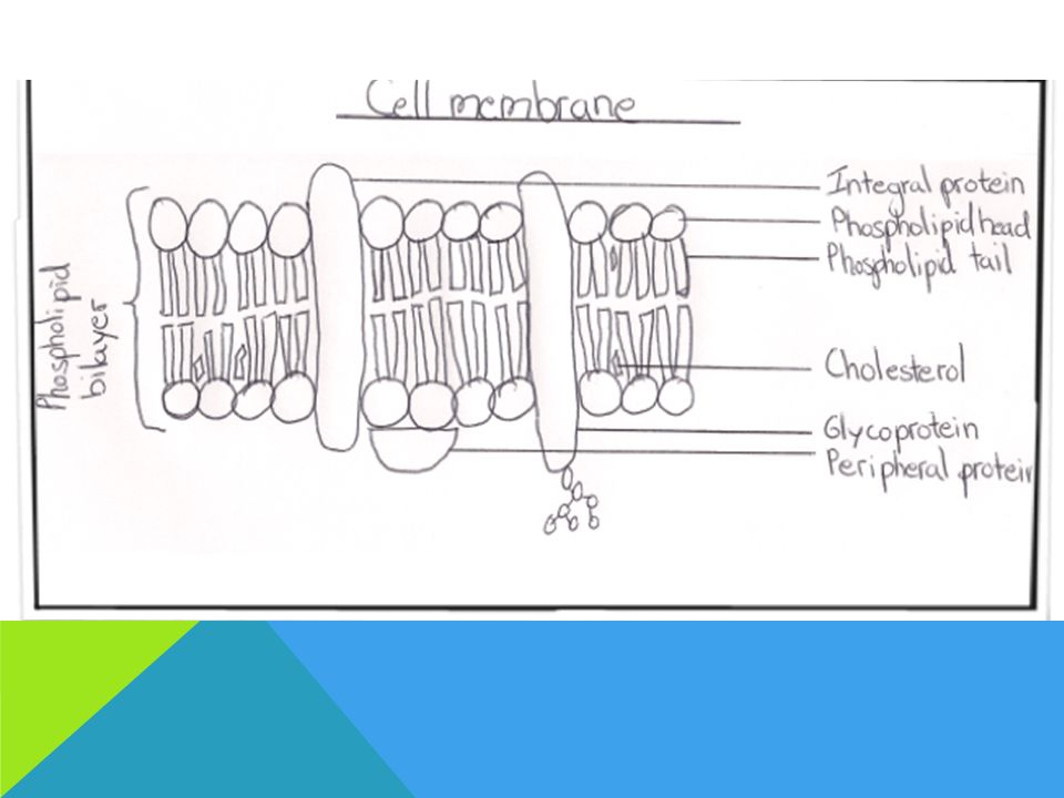
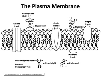
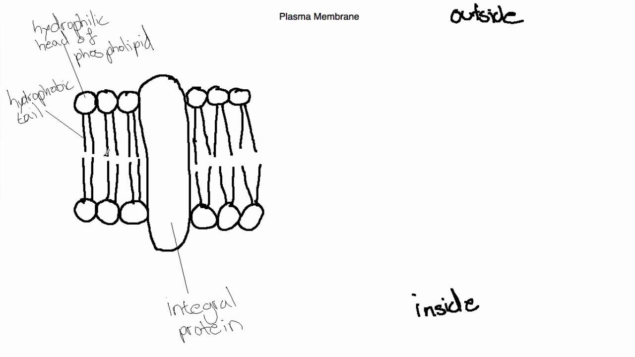
Labeled Plant Cell With Diagrams | Science Trends The parts of a plant cell include the cell wall, the cell membrane, the cytoskeleton or cytoplasm, the nucleus, the Golgi body, the mitochondria, the peroxisome's, the vacuoles, ribosomes, and the endoplasmic reticulum. Parts Of A Plant Cell The Cell Wall Let's start from the outside and work our way inwards. Solved Match the cell membrane structure or its function - Chegg Expert Answer Answer - phospoliods BILAYER . Labelled bilayer membrane . the hydrophilic part. Of the phospholipid bilayer are attract … View the full answer Transcribed image text: Match the cell membrane structure or its function with the correct letter from the diagram Label the hydrophobic and hydrophobic portions of the phospholipids. Simple Columnar Epithelium: A Labeled Diagram and Functions These form a brush border. They also increase the absorptive surface area of these cells. On a concluding note, simple columnar epithelium has two primary functions of absorption and secretion. In the small intestine, it facilitates the absorption of nutrients. It also secretes mucus, which helps to lubricate, moisten, and protect the surface. Plasma Membrane Function, Structure & Diagram - Study.com 2. property of the plasma membrane that allows some substances into the cell and keeps others out 4. main structural component of the plasma membrane 6. nonpolar part of a phospholipid 11. protein...
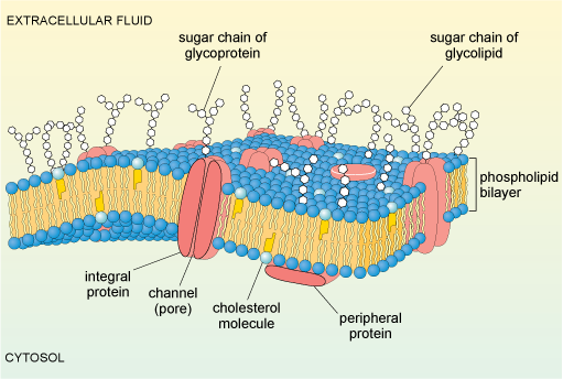
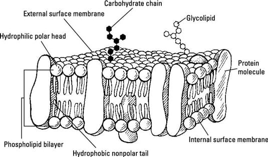
Plant Cell: Diagram, Types and Functions - Embibe Exams Xylem is a tissue that is formed of four different types of cells, i.e. tracheids, xylem vessels, xylem fibres and xylem parenchyma. They are the transport cells in vascular plants. They help in the transport of water and minerals from the roots to the leaves and other parts of the plants. The movement of water is unidirectional. Phloem highqualityanimalswallpaper.pages.dev highqualityanimalswallpaper.pages.dev File:Cell membrane detailed diagram id.svg - Wikimedia Original upload log []. This image is a derivative work of the following images: File:Cell_membrane_detailed_diagram_en.svg licensed with PD-user . 2009-02-23T18:08:26Z Bibi Saint-Pol 877x361 (487132 Bytes) {{Information |Description= {{en|The cell membrane, also called the plasma membrane or plasmalemma, is a semipermeable lipid bilayer common to all living cells. Cell membrane with labeled educational structure scheme vector ... 2. Editable Vector .EPS-10 file. 3. High-resolution JPG image. Use for everything except reselling item itself. Description: Cell membrane with labeled educational structure scheme vector illustration. Anatomical closeup drawing with cross section element. Carbohydrate, globular protein or cholesterol location visualization.
2.4.1 Draw and label a diagram to show the structure of ...
Labeled Diagram Of Cell Membrane : Electron Micrograph The nucleus and mitochondria are two examples. Copy of labeling cell membrane labelled diagram. Some of the major parts of the plasma membrane are : Phospholipid bilayer · phospholipid bilayer ; It supports and helps maintain a cell's shape. 1)cell membrane 2)vacuole 3)nucleus 4)endoplasmic reticulum 5)mitochondria 6)golgi body.

Membranes
2.4.1 Draw and label a diagram to show the structure of membranes Apr 25, 2012 ... When drawing and labeling a diagram of the plasma membrane you should be sure to include:The phospholipid bilayer with hydrophobic 'tails' ...
Cell Membrane: Definition, Structure, & Functions with Diagram
Structure of Membrane in Cells (With Diagram) - Biology Discussion In Gram-positive bacteria, the cell wall (Fig. 2.11) is (30-100 nm) thick outside the plasma membrane. The cell wall consists of peptidoglycan which is a polysaccharide- peptide complex. The polysaccharide complex of the adjacent chains are joined together by peptide bridges containing different kinds of amino acids like D and L-alanine, D ...
Biology Diagram Show Structure Cell Membrane Stock Vector ...
Draw and label a simple line diagram of a cell membrane. Include ... The outer covering of the body cells, which maintains homeostatic condition between inside and outside of the cell is called cell membrane. It is made up of ...
BIOLOGY 11 IB 2.4: MEMBRANES. ASSESSMENT STATEMENTS 2.4.1Draw ...
Labeling cell membrane - Teaching resources - Wordwall Quiz: Simple Cell Membrane Labeling Labelled diagram. by Cdesimone · Bio B - Cell Membrane Structure labeling Labelled diagram. by Karyn2.
Cell Membrane Structure (1.3)
Cell Organelles- Definition, Structure, Functions, Diagram - Microbe Notes A cell wall is multilayered with a middle lamina, a primary cell wall, and a secondary cell wall. The middle lamina contains polysaccharides that provide adhesion and allow binding of the cells to one another. After the middle lamina is the primary cell wall which is composed of cellulose.
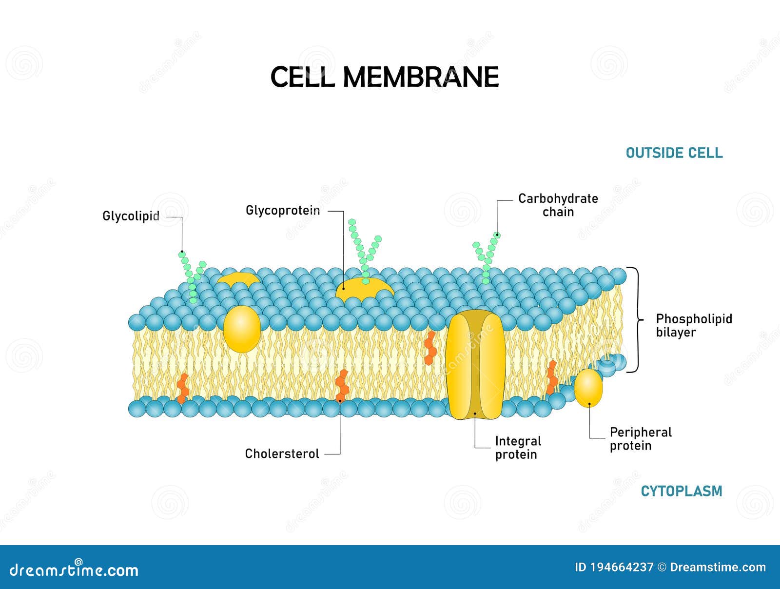
Diagram of Cell Membrane,phospholipid Bilayers Structure ...
Human Cell Diagram, Parts, Pictures, Structure and Functions Diagram of the human cell illustrating the different parts of the cell. Cell Membrane. The cell membrane is the outer coating of the cell and contains the cytoplasm, substances within it and the organelle. It is a double-layered membrane composed of proteins and lipids. The lipid molecules on the outer and inner part (lipid bilayer) allow it to ...

File:Cell membrane detailed diagram 3.svg - Wikimedia Commons
Cell Membrane (Plasma Membrane) - Genome.gov The cell membrane, also called the plasma membrane, is found in all cells and separates the interior of the cell from the outside environment. The cell membrane consists of a lipid bilayer that is semipermeable. The cell membrane regulates the transport of materials entering and exiting the cell. Narration 00:00 …
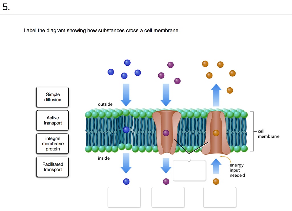
SOLVED: 5 Label the diagram showing how substances cross a ...
Structure of the plasma membrane (article) | Khan Academy The principal components of the plasma membrane are lipids (phospholipids and cholesterol), proteins, and carbohydrate groups that are attached to some of the lipids and proteins. A phospholipid is a lipid made of glycerol, two fatty acid tails, and a phosphate-linked head group. Biological membranes usually involve two layers of phospholipids ...

Biology Cell Membrane Diagram | Quizlet
Basic Cell Membrane Label - Labelled diagram - Wordwall Integral Protein (channel), Peripheral Protein, Phosphate, Lipid, Hydrophilic, Hydrophobic, Glycoprotein.
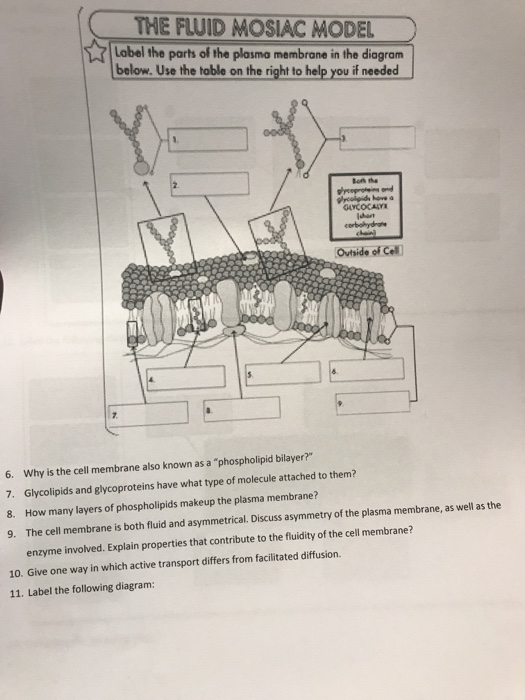
Solved THE FLUID MOSIAC MODEL Label the parts of the plasmo ...
PDF Human Cell Diagram, Parts, Pictures, Structure and Functions The cell membraneis the outer coating of the cell and contains the cytoplasm, substances within it and the organelle. It is a double-layered membrane composed of proteins and lipids. The lipid molecules on the outer and inner part (lipid bilayer) allow it to selectively transport substances in and out of the cell. Endoplasmic Reticulum

Animal Cell- Definition, Structure, Parts, Functions, Labeled ...
Cell Membrane Function and Structure - ThoughtCo The cell membrane (plasma membrane) is a thin semi-permeable membrane that surrounds the cytoplasm of a cell. Its function is to protect the integrity of the interior of the cell by allowing certain substances into the cell while keeping other substances out.
Cell membrane stock vector. Illustration of cellular - 98137872
Animal Cells: Labelled Diagram, Definitions, and Structure - Research Tweet The endoplasmic reticulum (s) are organelles that create a network of membranes that transport substances around the cell. They have phospholipid bilayers. There are two types of ER: the rough ER, and the smooth ER. The rough endoplasmic reticulum is rough because it has ribosomes (which is explained below) attached to it.

Cell membrane - Wikipedia
Cell Diagrams with Labelling Activity - Learnful The cell structure illustrations for these diagrams were generated in BioRender. Both diagrams feature a drag-and-drop labelling activity created with H5P here ...
Cell membrane or cytoplasmic membrane microscopic structure outline diagram
Labeling a cell membrane Diagram | Quizlet Labeling a cell membrane Diagram | Quizlet Labeling a cell membrane 5.0 (8 reviews) + − Learn Test Match Created by EGSchumacher Terms in this set (9) Hydrophobic tail ... Hydrophilic head ... Channel Protein (integral protein) ... Glycolipid ... Cholesterol ... Phospholipid Bilayer ... Glycoprotein ... Peripheral Protein ... Carbohydrate Chain ...

Fluid Mosaic Model Diagram | The Fluid-Mosaic Model of the ...
Label Cell Membrane Diagram - Quizlet Start studying Label Cell Membrane. Learn vocabulary, terms, and more with flashcards, games, and other study tools.

Draw and label a simple line diagram of a cell membrane ...
Schematic Diagram of a Cell Membrane - Pinterest Honors Biology @ Lawrenceville. Today we discussed the details of the cell membrane. What is it made out of?
Animal Cell Membrane - Interactive DiagramkidCourses.com
CELL MEMBRANE LABEL Diagram | Quizlet Practice labeling the parts of the cell membrane Terms in this set (6) Channel Protein hole or tunnel that particles may pass through to go in / out of cell Marker protein identifies or labels the cell Receptor protein receives information Heads part of the phospholipid that loves water (hydrophili) - points to the most outside and inside of cell
Cell Wall and Cell Membrane- Structure, Functions and Differences
Cell: Structure and Functions (With Diagram) - Biology Discussion Eukaryotic Cells: 1. Eukaryotes are sophisticated cells with a well defined nucleus and cell organelles. 2. The cells are comparatively larger in size (10-100 μm). 3. Unicellular to multicellular in nature and evolved ~1 billion years ago. 4. The cell membrane is semipermeable and flexible. 5. These cells reproduce both asexually and sexually.
:max_bytes(150000):strip_icc()/plasma_membrane-58a617c53df78c345b5efb37.jpg)
Cell Membrane Function and Structure
Cell Membrane - The Definitive Guide | Biology Dictionary The cell membrane, also known as the plasma membrane, is a double layer of lipids and proteins that surrounds a cell. It separates the cytoplasm (the contents of the cell) from the external environment. It is a feature of all cells, both prokaryotic and eukaryotic. a 3D diagram of the cell membrane Function of the Cell Membrane

Diagram With Plasma Membrane Stock Illustration - Download ...
Diagram of a cell membrane with labels - nist.gov Essential Biological FunctionsImmune response, Cell metabolism, Neurotransmission, Photosynthesis, Cell adherence, Cell growth and differentiationPotential Commercial ApplicationsDrug response monitoring, Chemical manufacturing, Biosensing, Energy conversion, Tissue engineering ... Diagram of a cell membrane with labels. Appears In. Biology in ...
A tour of the cell: View as single page

1. Labeling a cell membrane Diagram | Quizlet

Cell Membrane Functions, Structure and Diagram
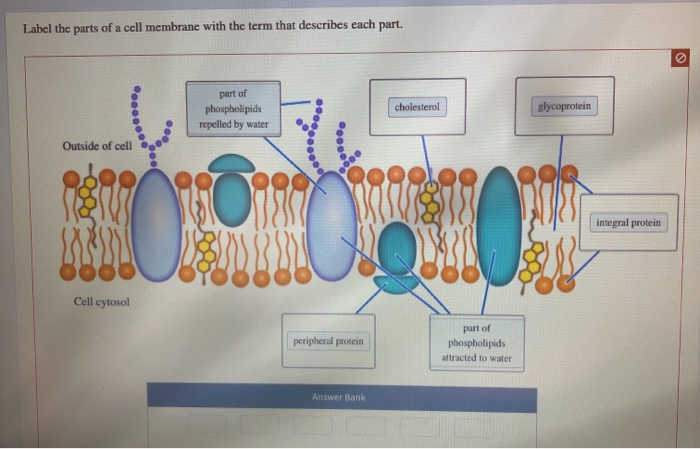
Solved Label the parts of a cell membrane with the term that ...

Biology Pictures: Cell Membrane Structure without labels ...
Labeling cell membrane - Teaching resources
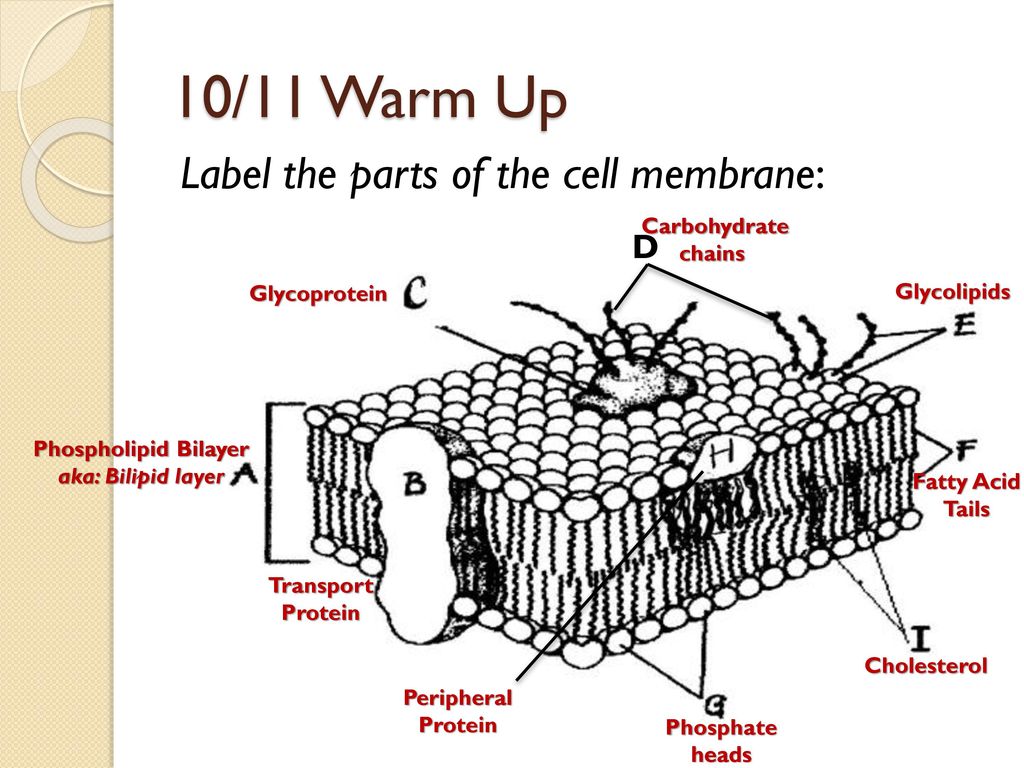
10/11 Warm Up Label the parts of the cell membrane: D ...
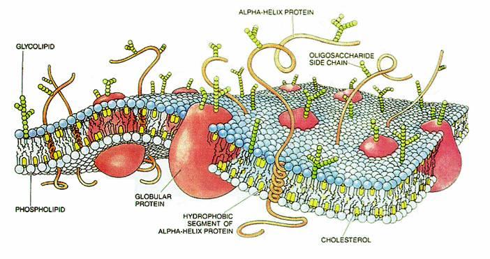
Diagram of a cell membrane with labels
Human cell membrane structure illustration Stock Vector Image ...

help me label this cell membrane pls - Brainly.com
Cell Membrane Structure (1.3)
Membranes I | Biology | Visionlearning
Plasma Membrane / Fluid Mosaic Diagram
The Cell Membrane: Diffusion, Osmosis, and Active Transport ...
IB Biology Topic 2.4.1 Draw and Label the Plasma Membrane
:max_bytes(150000):strip_icc()/cell-membrane-373364_final-5b5f300546e0fb008271ce52.png)
Cell Membrane Function and Structure
Plasma Membrane Markers: Novus Biologicals
Cell Membrane Stock Illustrations – 5,870 Cell Membrane Stock ...

Cell Membrane Quiz: Membrane Structure And Function Quiz ...
Cellular organelles and their functions | Kenhub
Post a Comment for "41 cell membrane diagram with labels"